What’s New in CT Systems and Scanner Technology in 2024
“The art of medicine consists in amusing the patient while nature cures the disease.” – Voltaire
If you’re wondering how CT systems have advanced and what new features define the latest scanners, you’re in the right place. From enhanced image resolution to AI integration, discover the pivotal innovations in CT technology that are setting new standards in medical imaging in 2024.
Key Takeaways
-
Even with new technological advancements in the medical imaging field, we find many organizations that are paying for more features than they need. At Mansa Medical, we offer low-cost options that get you exactly what you need for your patients without paying the extra price for largely unnecessary features.
-
Innovations in CT technology aim to reduce radiation dose through automatic exposure control and real-time dose monitoring, prioritize safety with the ALARA principle, and leverage photon-counting CT scanners like Siemens’ NAEOTOM Alpha for enhanced image quality and safety.
-
Artificial intelligence is increasingly integrated into CT technology, automating routine tasks, improving diagnostic accuracy, and streamlining workflows, as exemplified by systems like the Philips CT 3500, which optimizes image reconstruction and enhances image quality.
CT Systems: An In-Depth Look
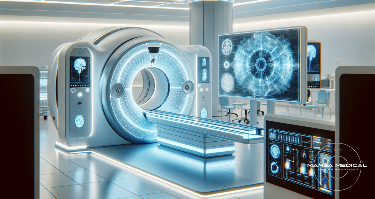
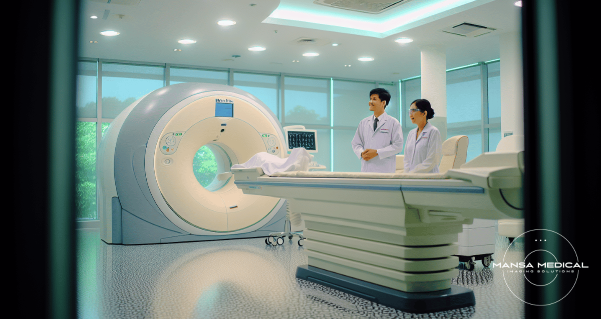
Known as computed tomography systems or computed tomography CT, these systems have revolutionized medical imaging with their detailed body views surpassing conventional X-rays. They use a rotating X-ray source and detectors to capture precise cross-sectional images of the body through computed axial tomography. The X-ray source in a CT system emits narrow beams of X-rays that pass through the patient’s body, creating an X-ray image. As these rays pass through, detectors within the CT system capture them.
The process of obtaining a computed axial tomography scan, also known as CT scan, involves the following steps:
-
The patient is positioned on a table that slides into the CT scanner.
-
X-ray beams are emitted from multiple angles around the patient’s body.
-
The captured X-rays are then transmitted to a computer, which reconstructs them into detailed images.
-
These images provided by CT systems offer greater clarity and more detailed information than conventional X-ray images, allowing for an accurate diagnosis of various medical conditions.
-
CT scans are used not only for detecting bone fractures but also for viewing soft tissues and blood vessels.
-
Contrast dye, also known as contrast material, can be used to highlight certain areas, making it easier to observe internal injuries or abnormalities.
As you are aware, CT scanners, also known as CAT scan machines, have been instrumental in examining internal organs, soft tissues, and solid organs [1]. A CT imaging system generates a detailed cross-sectional view that provides more detailed information than traditional chest X-ray or spiral CT. One of the key elements of the CT system is the CT fan beam, which is responsible for emitting the narrow beams of X-rays.
These CT images are instrumental in diagnosing diseases, monitoring therapy, and planning surgeries. For instance, a CT angiogram can give detailed images of blood vessels in the brain, aiding in the diagnosis of vascular diseases. With a CT examination, doctors can visualize the entire structure of the patient’s digestive system, from the esophagus to the rectum, assisting in the diagnosis and treatment of digestive diseases. The detailed images provided by CT scans make it an invaluable tool in modern healthcare.
Innovations in CT Technology: What to Expect in 2024?

As established, CT systems play a crucial role in medical imaging. Yet, akin to all technologies, their evolution persists. The year 2024 is already witnessing multiple pivotal innovations in CT technology, enhancing image quality, reducing radiation dose, and improving accessibility.
These innovations include:
-
The introduction of photon-counting technology
-
Improvements in image reconstruction techniques
-
The emergence of mobile CT systems
-
Enhancements in resolution and speed
-
The integration of artificial intelligence
Each of these advancements brings its own unique benefits, and collectively, they are transforming the way we use CT technology. Let’s go over them in detail.
Reduced Radiation Dose
Radiation exposure, a potential risk posed by any imaging form using ionizing radiation like CT scans, remains a key concern in developing cancer [2]. However, modern CT scanners have made significant strides in minimizing this risk, focusing particularly on reducing the radiation dose.
Automatic exposure control (AEC) systems in modern CT scanners adjust the radiation based on the patient’s size and shape, minimizing unnecessary exposure. This is particularly important when performing pediatric imaging, where the ALARA principle – ‘As Low As Reasonably Achievable’ – is crucial for safety.
Photon-counting CT scanners have been particularly instrumental in this regard. These scanners enhance geometrical dose efficiency, which leads to reduced radiation doses for patients. This technology, introduced by Siemens Healthineers in the form of the NAEOTOM Alpha, has marked a significant advancement in CT technology.
Additionally, real-time dose monitoring in CT scanners enables on-the-fly adjustments to radiation levels, ensuring patient radiation safety throughout. This feature allows the scanner to adapt to the specific needs of the patient without compromising image quality.
The focus on reducing radiation dose is not limited to the technology alone. Optimizing CT facility quality assurance involves using protocols that apply the lowest radiation dose possible while maintaining image quality. This approach ensures that the benefits of CT scans can be achieved with the least possible risk.
Improved Image Reconstruction Techniques
Besides reducing radiation dose, CT scan machine technology has made remarkable progress in enhancing image reconstruction techniques. Image reconstruction is the process by which the data captured by the CT scanner is converted into cross-sectional images. This process is integral to the quality of the CT images and, consequently, the accuracy of the diagnosis [3].
Historically, filtered backprojection (FBP) was the predominant reconstruction method. However, new iterative reconstruction techniques are now replacing it to improve image quality, particularly at reduced radiation doses. One such technique is model-based iterative reconstruction (MBIR). MBIR offers the following benefits:
-
Significantly improves spatial resolution
-
Reduces noise
-
Lower image noise and higher resolution than FBP
-
Accurate diagnosis
-
Better patient outcomes
Artificial intelligence (AI) and deep learning have also found their way into CT image reconstruction. The integration of these technologies shows substantial promise for:
-
Enhanced image quality
-
Potential for additional radiation dose reductions
-
Improved diagnostic accuracy
-
Increased efficiency
-
Potential reduction in healthcare costs.
Physical factors are now more accurately incorporated into image reconstruction, leading to improvements in image quality. In addition, advanced algorithms like ADMIRE reduce artifacts, and noise reduction techniques fine-tune the balance between high-contrast resolution and low-contrast detectability.
Photon-Counting
Of all the innovations in CT technology, the emergence of photon-counting CT arguably stands as the most revolutionary. This technology streamlines the conversion of X-ray photons into electrical signals, a process that was traditionally a two-step conversion involving visible light. This change has brought about significant improvements in image quality and safety.
Traditionally, X-ray photons were first converted into visible light, which was then converted into an electrical signal. However, the introduction of the NAEOTOM Alpha, a photon-counting CT by Siemens Healthineers, has changed the game. This technology directly converts X-ray photons into electrical signals, skipping the visible light conversion step.
As a result of this streamlined process, photon-counting CT offers several advantages:
-
Preserves all of the original X-ray information
-
Provides radiologists with high spatial resolution CT data
-
Improves contrast-to-noise ratio
-
Produces imagery with no electronic noise
-
Results in a lower radiation dose with intrinsic spectral information.
The introduction of photon-counting technology is truly a testament to the continuous evolution and advancement of CT technology. It underscores the relentless pursuit of improved patient safety, superior image quality, and enhanced diagnostic precision in the world of medical imaging.
Mobile CT Becomes More Common
The rise of mobile CT systems, such as those you can buy or rent at Mansa Medical, signifies another notable progression in CT technology. These systems have addressed some of the major challenges faced by healthcare providers, particularly in intensive care units (ICUs).
One of the biggest challenges in ICUs is:
-
The process of getting patients to the CT scanner
-
The transport of patients, especially those with head trauma, carries inherent risks and requires highly qualified team members
-
Ongoing staffing challenges that need to be addressed.
To mitigate these risks and challenges, many providers have turned to mobile CT systems. Mobile CT systems like the SOMATOM can be brought to the patient’s bedside, eliminating the need for patient transport. This not only reduces risks associated with patient transport but also addresses staffing challenges and provides a high-quality experience for patients and staff alike.
The advent of mobile CT systems is a testament to how innovations in CT technology are continually reshaping healthcare delivery. By bringing CT imaging directly to the patient, these mobile systems are revolutionizing patient care and have the potential to transform how CT scans are acquired in the future.
Enhanced Resolution and Speed
With the growth of computational power and the sophistication of software algorithms, enhancements in resolution and speed have become significant achievements in CT technology. These enhancements not only benefit patient comfort but also enable more precise diagnoses.
One of the most anticipated developments in CT technology is the improvement in resolution and speed. It is noted that the advancement in computational power and software algorithms have empowered CT scanners to produce even more detailed images in a shorter amount of time. This is particularly beneficial for conditions that require capturing dynamic processes within the body. The improved resolution allows for a more accurate depiction of these processes, enabling physicians to make more precise diagnoses.
Furthermore, the increased speed of these scans reduces the discomfort for the patient and can potentially reduce the time taken for diagnosis and treatment initiation. These advancements in resolution and speed highlight the continuous evolution of CT technology. With these ongoing improvements, CT scans are becoming more effective as a diagnostic tool, providing even more detailed images that enable healthcare professionals to deliver high-quality patient care.
Artificial Intelligence Integration
Artificial intelligence (AI) is poised to assume a central role in the future development of CT technology. With its ability to automate routine tasks and improve image interpretation, AI integration is transforming the way CT technology is used in medical imaging.
AI algorithms can assist radiologists in image interpretation. They also help by automating routine tasks such as organ segmentation, tumor detection, and anomaly identification. This not only improves diagnostic accuracy but also increases efficiency, potentially reducing healthcare costs.
An example of this integration is the Philips CT 3500, a new high-throughput CT system powered by AI. This system includes a range of image-reconstruction and workflow-enhancing features that help deliver consistent, fast, and high-quality images, which are crucial for confident diagnoses.
The integration of artificial intelligence in CT technology is a clear indication of how technology is reshaping the healthcare industry. It underscores a future where AI and machine learning will continue to play an increasingly important role in improving patient care and outcomes.
Benefits of Used MRI and CT Machines
Even though technology is always advancing in the imaging medical field, used equipment still brings many benefits and can be advantageous for medical facilities. Primarily, they offer significant cost savings compared to buying new equipment, allowing for more efficient budget allocation. Moreover, the acquisition process for used machines is often quicker, enabling faster implementation and improved patient care.
Used equipment often has a proven track record of reliability, as sellers typically refurbish and thoroughly test it to meet industry standards. The extended lifecycle of medical imaging equipment makes used machines a cost-effective option, providing access to advanced technology without the full expense associated with new equipment.
Additionally, some used machines may come with extra features, upgrades, or accessories, enhancing their capabilities. Avoiding the steeper initial depreciation associated with new equipment, used machines may experience slower depreciation over their remaining useful life.
Buying used equipment for smaller healthcare facilities with limited budgets can be more feasible than investing in new machines. This allows them to access advanced imaging technology without compromising on patient care. Furthermore, for facilities with specific and occasional imaging needs, used equipment is a cost-effective solution compared to the expense of a new machine.
Despite these benefits, careful assessment of the equipment’s condition, maintenance history, and specifications is essential before making a purchase. Working with reputable sellers and ensuring compliance with regulatory standards are crucial aspects of a successful acquisition.
Mansa Medical: Pioneering Technological Progress in MRI and CT Solutions, Including Mobile CT Rentals for Flexible Healthcare Access

Leading these technological advancements, Mansa Medical delivers innovative CT and MRI solutions. Specializing in multi-facility upgrades, machine installations, and service and project management, Mansa Medical is committed to providing equipment that is clean, safe, and reliable and showcases cutting-edge technology.
For those seeking temporary solutions, Mansa Medical offers mobile CT rentals, bringing the benefits of advanced CT technology directly to the patient’s bedside. Contact Mansa Medical today!
Full Summary
In the ever-evolving world of medical imaging, CT technology continues to make significant strides. From reducing radiation doses to improving image reconstruction techniques, from introducing photon-counting technology to the advent of mobile CT systems, and from enhancing resolution and speed to integrating artificial intelligence, each innovation brings its own unique benefits, and collectively, they are transforming the way we use CT technology.
As we navigate through this exciting era of technological advancements, companies like Mansa Medical are leading the way, providing innovative solutions that prioritize patient safety and care. These developments not only enhance our ability to diagnose and treat diseases but also underscore a future where technology continues to revolutionize healthcare.
Frequently Asked Questions
Do mobile CT machines exist?
Yes, Siemens Healthineers offers a mobile head CT scanner called SOMATOM, providing reliable imaging at a patient’s bedside. Transform the way you care for ICU patients suffering from acute and neurocritical conditions.
What is a CT used for?
A CT scan is used to obtain detailed images of the body, including bones, muscles, organs, and blood vessels, as well as for fluid or tissue biopsies and preparation for surgery or treatment.
What are the three major components of the CT system?
The three major components of the CT system are an X-ray tube, a gantry with a ring of X-ray-sensitive detectors, and a computer. These components work together to create images using the same physics principles as in conventional radiography.
Are there different types of CT machines?
Yes, there are different types of CT machines with slice counts, including 16, 32, 64, 128, 256, and 320-slice CT scanners, each suitable for different types of studies. It’s important to select a CT scanner that aligns with your specific needs and budget, as well as patient flow targets.
How does a CT system work?
A CT system works by using a rotating X-ray source and detectors to capture detailed cross-sectional images of the body, providing clearer and more detailed information than conventional X-rays. This helps in accurate diagnosis and treatment planning.
